Why This Comparison Matters
Many people encountering GDV technology for the first time assume it's "just Kirlian photography with a new name." This article explains the precise technical differences — and why those differences are the reason GDV has 400+ published studies while traditional Kirlian photography's scientific credibility remained limited. We also address both technologies' known limitations honestly.
In This Guide
The Same Physics, Different Execution
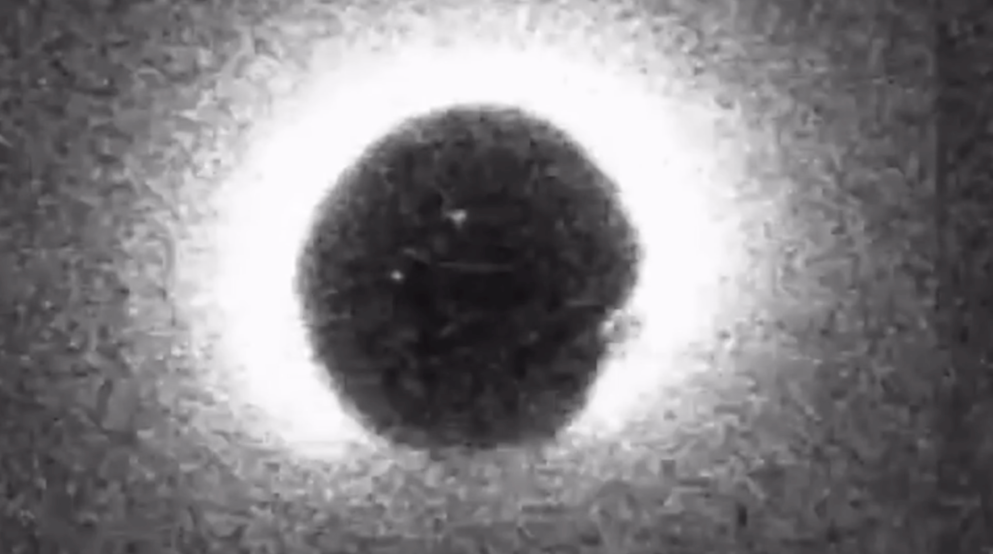
Both Kirlian photography and GDV imaging rely on the same underlying physical phenomenon: gas discharge. When a high-voltage electrical field is applied to an object on a conductive surface, the gas molecules surrounding that object become ionized, and the resulting electron cascade emits visible light — a corona discharge.
This phenomenon was first documented by Georg Christoph Lichtenberg in 1777 and has been explored by Nikola Tesla (1891), Narkevich-Yodko (1893), and Semyon Kirlian (1939). The physics hasn't changed. What changed is how we capture, control, and analyze the resulting images.
The core equation
High-voltage pulse + living tissue + gas = visible corona discharge
This is true for both Kirlian and GDV. Everything else is engineering.
Head-to-Head: 10 Technical Differences
| Feature | Kirlian Photography 1939–1990s |
GDV / Bio-Well 1995–present |
|---|---|---|
| 1. Capture medium | Photographic film (chemical development required) | CCD digital sensor (instant capture) |
| 2. Output format | Visual image only — qualitative | Digital data + image — quantifiable (area, intensity, entropy, fractality) |
| 3. Calibration | None — each setup had unique parameters | Built-in calibration unit, standardized protocol |
| 4. Reproducibility | Low — affected by 200+ variables (moisture, pressure, voltage, humidity, etc.) | Improved — controlled parameters and filters, though some variability persists |
| 5. Interpretation | Subjective — practitioner visually reads the image | Algorithmic — software computes parameters from pixel data |
| 6. Organ mapping | Mandel's EEA system (1986) — manual, practitioner-dependent | Computerized meridian mapping with sector analysis per fingertip |
| 7. Speed | Minutes per finger + darkroom development time | ~6 seconds per finger, instant software analysis |
| 8. Before/after | Possible but impractical (two film sets, darkroom comparison) | Built-in software overlay — real-time comparison |
| 9. Reference database | None | 1M+ scans (manufacturer-stated) |
| 10. Published studies | Limited, with reliability concerns noted in multiple reviews | 400+ papers (per IUMAB database), though methodological rigor varies |
Sources for Kirlian limitations: Pehek, J.O. et al. "Image Modulation in Corona Discharge Photography." Science. 1976;194(4262):263-270 — identified moisture as the principal determinant of corona discharge variations. Overview → · For GDV evidence base: IUMAB research database →
Interactive: Myths vs. Documented Facts
Confusion between Kirlian photography and modern GDV has created persistent myths. Tap each claim to see what the published research actually supports.
Why Kirlian Photography Hit a Scientific Wall
Traditional Kirlian photography produced striking, beautiful images. But its scientific credibility was undermined by several documented problems that multiple research groups identified over decades:
Moisture dominated the signal
The 1976 Science paper by Pehek, Kyler, and Faust concluded that moisture content on the skin surface was the principal determinant of corona discharge appearance — meaning what many interpreted as "energy" was largely sweat.
Too many uncontrolled variables
Researchers identified as many as 200 factors that could influence the image: voltage, frequency, pressure, exposure time, humidity, temperature, film type, development chemistry, and more. Without standardization, no two setups produced comparable results.
Subjective interpretation
Without computer analysis, all interpretation was visual and practitioner-dependent. Different practitioners could read the same image differently — a problem one researcher compared to a "Rorschach process."
No statistical validation
Multiple reviews noted a lack of systematic clinical studies with adequate sample sizes. Without numbers to analyze statistically, the images remained interesting but scientifically unverifiable.
Sources: Pehek, Kyler, Faust. Science 194(4262):263-270, 1976. · Eidson et al., Drexel University, 1976/1978 (U.S. Department of Defense). · Wikipedia: Kirlian photography (overview of scientific critiques) →
What GDV Solved — and What It Didn't
When Korotkov developed GDV in 1995, he specifically targeted the problems that had undermined Kirlian photography's credibility. Here's what changed — and what remains a work in progress:
Where the Technology Stands Today
Bio-Well 3.0 — the latest generation of GDV technology — represents the current state of the art. It incorporates all of the advances described above: digital CCD capture, built-in calibration, disposable filters, standardized protocols, and software analytics drawing on a large reference database.
Over 5,000 practitioners in 70+ countries currently use Bio-Well, and the IUMAB research database continues to grow. The technology has come an extraordinary distance from Semyon Kirlian's first accidental corona discharge photographs in 1939 — but like all scientific instruments, it continues to be refined, tested, and improved.
See the difference for yourself
Watch a free demo to see what modern GDV technology looks like in practice.
Sources Cited in This Article
- Pehek, J.O., Kyler, H.J., Faust, D.L. "Image Modulation in Corona Discharge Photography." Science. 1976;194(4262):263-270. Context →
- "Kirlian photography." Wikipedia. Comprehensive overview of scientific research and critiques. Article →
- Korotkov, K. "Review of EPI papers on medicine and psychophysiology 2008–2018." Int J Complement Alt Med. 2018;11(6). DOI →
- IUMAB published research database. iumab.club →
- "Correlation of EPI Parameters With Fasting Blood Sugar." J Evidence-Based CAM, 2016. PDF →
- "Causal Relationships Between GDV Parameters and Leukocytogram." J Education, Health and Sport, 2021. PDF →
- Cioca, G. et al. "GDV and HRV Correlation." 2004. PDF →
- Bio-Well 3.0 product specifications. bio-well.com →
- Bio-Well Science page (GDV history timeline). bio-well.com/pages/science →
- Eidson, W. et al. Drexel University study, 1976/1978. Published via U.S. Department of Defense documentation.
- Blacklock, N.F. "Imaging the Bio-Field: Kirlian Photography and the Eight Extraordinary Meridians." GDV-meridian correlation study.
- Bio-Well official biography of Pr. Korotkov. bio-well.com →

Share:
Biofield healing: what the research says about energy-based therapies
Bio-Well 3.0: what's new in the latest GDV device